Medical Services
Providing CBCT examination
In a workplace Department of Stomatology, General Teaching Hospital and First Faculty of Medicine it is possible to take three-dimensional X-ray examination of teeth and bones of the head with Cone Beam Computed Tomography (CBCT).
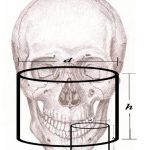
It is possible to selectively select the size of the display area, see the diagram Field of View (FOV)
- from 5×5 cm high resolution for endodontic and isolated implantology purposes
- over 8×8 and 10×10 cm for multiple implantations in both jaws
- after 17×13,5 cm for maximum facial skeleton display
Forms
ON-LINE ordering of CBCT examination (completed request with you)
The patient will receive a CD with the examination, including the original browser, immediately after the examination. We do not provide another output format!
To view the 3D model, it is necessary to run the software with installation on a PC (see "Instructions for installing the CBCT scan viewer").
Other documents
- Instructions for installing CBCT scans (PDF)
- An exemplary CBCT scan of the facial skeleton (ZIPPER)
- Installer for viewing CBCT scans (EXE)
Manuals for controlling CBCT scans on your computer (English webinars)
Introduction to 3D CBCT Diagnostics
- FOV 17 × 13 and FOV 5 × 5 on lbi
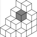
- voxel
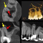
- yellow arrow - first channel (MB1), red arrow - second channel (MB2)
CBCT - Cone Beam Computed Tomography. An X-ray imaging that captures the selected tissue volume in all planes to provide a spatial overview and diagnostic information from them.
FOV - Field of View (Volume of investigated area). It is a cylinder with a given diameter (first value) and height (second value) given in centimeters, eg 5 × 5, 10 × 5, etc. It is necessary to know the size of the structure (in centimeters) you want to display ( eg a single tooth, maxila, both jaws, etc.) and choose a similar adequate FOV size. It is also necessary to select the FOV localization targeted to cover the given structure (eg upper left dental sextant, left rock bone, etc.)
Voxel - Volumetric Picture Element. The smallest volume subunit of the 3D object model. The computer model of an object can be divided into small cubes (voxels). These cubes (voxels) have a given dimension of the sides reported for CBCT in micrometers. The smaller their size, the greater the resolution of the 3D model, which allows for greater detail.
DICOM Viewer - A dedicated computer program is used to view 3D volumes. There are universal browsers that can open 3D DICOM volumes from most CBCT devices. Most CBCT devices can export their 3D volumes with the included browser and variously limited features over the full version.
The list of features that should not be missing in the 3D volume viewer is as follows:
- scrolling through any section in all planes
- reconstruction of 3D volume model with possibility of filtration of x-ray contrast layers
- any adjustment of the section plane (essential function for endodontic diagnosis)
- measuring lengths and angles
- virtual implantation


